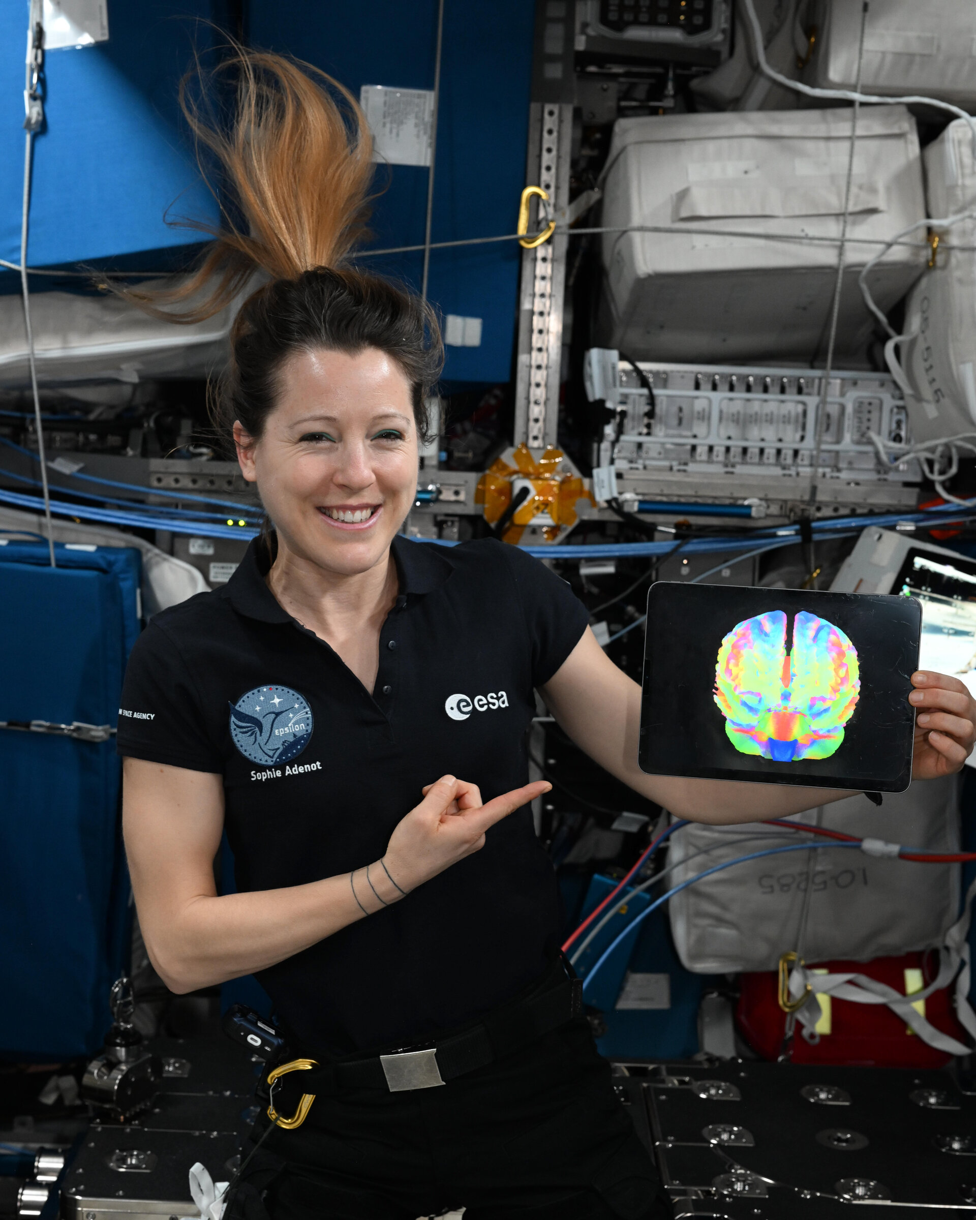
Coming home does not end a spaceflight experiment. For researchers studying the brain, landing starts a second, time-sensitive observation window: the return to gravity. An ESA update published October 30, 2026 describes that work following Sophie Adenot’s 237-day mission.
Adenot underwent three MRI sessions at DLR’s :envihab facility in Cologne within her first two weeks back. Another scan is planned six months after her return. The aim is to measure the course of readaptation, rather than assume that all changes reverse at the same rate.
Different scans answer different questions
Brain-DTI combines anatomical imaging with diffusion measurements, which track water movement through tissue, and resting-state functional MRI, which uses blood-oxygenation signals to investigate communication between brain regions. Together, these approaches can examine fluid redistribution, brain position and neural pathways. ESA says the project, which began collecting scans in 2013, now includes around 50 astronauts and cosmonauts.
This is an account of ongoing measurements, not a published diagnosis of Adenot or a final result from her recovery scans. That distinction matters whenever an astronaut’s photograph is used to illustrate a health investigation.
Why the time between flights matters
A separate 2023 study in Scientific Reports helps explain why recovery timing is scientifically important. Researchers compared preflight and postflight MRI data from 30 astronauts whose missions ranged from roughly two weeks to a year. Longer missions were associated with greater expansion of some fluid-filled brain ventricles, with much of that expansion occurring during the first six months.
The team also found an association with the interval between missions. Astronauts with less than three years between flights showed little or no additional enlargement in certain ventricles. The authors interpreted this as potentially reflecting incomplete recovery of their capacity to accommodate another flight—not proof that frequent flyers were unaffected.
Those findings do not establish a universal three-year recovery deadline. The study examined structural measures in a limited astronaut sample; it did not turn every MRI change into evidence of injury, nor did it show that every person follows the same trajectory. Applying its group results to one named crew member would go beyond the evidence.
The connection to vision
NASA’s overview of spaceflight-associated neuro-ocular syndrome, or SANS, describes changes including optic-nerve swelling and flattening at the back of the eye. Headward movement of fluids in weightlessness is a major focus of investigation. NASA says the long-term health consequences remain under study, with outcomes ranging from mild changes to clinically significant effects.
For future missions, the useful question is therefore more precise than whether space “changes the brain.” Researchers need to establish which changes represent adaptation, which affect function, and how quickly each resolves. Repeated scans paired with clinical observations can help separate those questions. A single before-and-after image cannot.
Featured photograph: Sophie Adenot holding a Brain-DTI scan. ©ESA/NASA, used for editorial reporting under the ESA Standard Licence.
Leave a Reply